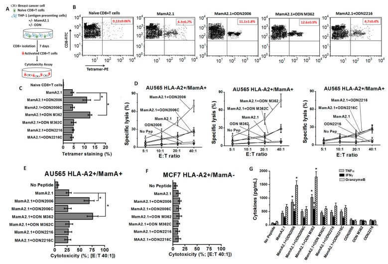
Figure 1.
Activation of CD8+T lymphocytes (CTLs) following antigen presentation by THP1 cells and stimulation by MamA2.1 peptide with and without ODNs. (A) Schematic of the experimental design. (B,C) MamA2.1 tetramer staining of the CTLs. The naïve CD8+T cells were stimulated by THP-1 cells pre-treated with either the MamA2.1 peptide alone or in combination with various ODNs. (D) Cytotoxicity of CTLs activated under various treatment conditions (mentioned in text) on AU565 breast cancer cells at effector to target (E:T) ratios (from 5:1 to 40:1). (E,F) Comparative cytotoxic efficiency of CTLs following various stimulations at E:T ratio 40:1 on AU565 (HLA-A2+/MamA+) and MCF7 (HLA-A2+/MamA−) breast cancer cell lines. (G) Cytokines analyzed from the whole cell lysate of CTLs. All experiments were performed in four independent replicates, and data expressed as mean ± SD, p < 0.05 compared with MamA2.1 peptide treatment alone. MamA: Mammaglobin-A; ODN: oligodeoxynucleotides.

