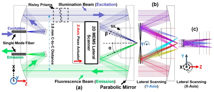
Figure 1.
Ray-tracing simulation of the optical design for the MEMS-based intraoperative near-infrared (NIR) confocal microscope’s scan-head (outer diameter (OD): 5.5-mm package). (a) Schematic drawing of the scan-head, such as the collimating, focusing, and scanning in the post-objective dual-axis confocal architecture aimed for 3D NIR fluorescence imaging, demonstrating the geometric requirements for the MEMS scanner; a pair of tiny Risley prisms were used for precise alignment; single-mode fibers (S630HP, numerical aperture (NA) = 0.12) were used for delivering and collecting light beams; SIL: solid immersion lens, β: free-space numerical aperture of the individual beams, and α: the intersection half-angle of the beams. (b) Lateral scanning around the Y-axis of the micro-mirror. (c) Lateral scanning around the X-axis of the micro-mirror.

