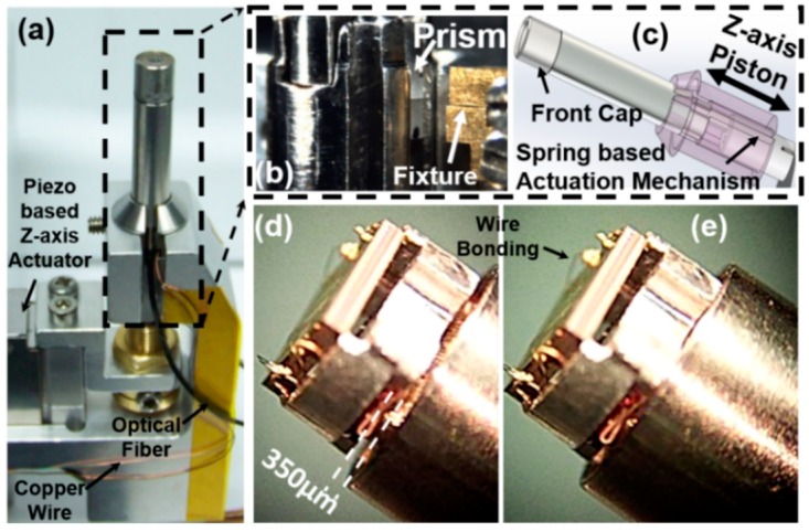
Figure 8.
Integration of the 2D resonant MEMS scanner in the miniaturized fiber-based intraoperative NIR fluorescence confocal microscope. (a) Stereomicroscopic image of the microscope; a piezoelectrical Z-axis actuator (P-601.4SL) was used for axial scanning (DC stacking mode, or axial scanning up to 5 Hz with a 350-µm range); (b) zoom-in view of the microscope’s distal end; Risley prisms were used for aligning the beams precisely to be parallel; additional metal alignment fixtures; (c) schematic drawing of the scan-head’s distal end and the spring-based Z-axis piston mode actuation mechanism; (d,e) zoom-in view of the Z-axis movement of the MEMS scanner holder stage with 350-µm movement; the front cap has been taken off.

