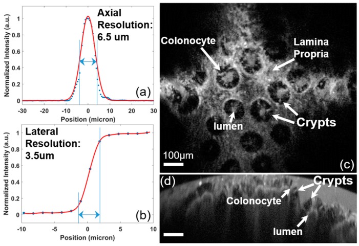
Figure 10.
Fluorescence imaging results of the NIR fluorescence intraoperative confocal microscope. (a) Lateral and (b) axial resolution at the wavelength of 785 nm (focus at 150 µm out of the SIL) by measuring the FWHM in the reflective mode; ex vivo fluorescence images of human colon tissue specimens demonstrated “histology-like” imaging with a large field-of-view (up to 1000 µm), in both the (c) en face horizontal cross-sectional image at the 150-µm depth and (d) vertical cross-sectional image; crypts, colonocytes, and lumen have been visualized with cellular resolutions, scale bar: 100 µm.

