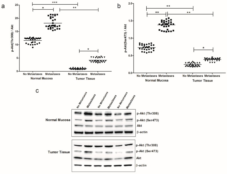
Figure 4.
(a) p-Akt (Thr308)/Akt ratio protein values, detected in intestinal tissue of no metastases (n = 29 patients) and with metastases patients (n = 30 patients), in both normal mucosa and tumor tissue. (b) p-Akt (Ser473)/Akt ratio protein values detected in intestinal tissue of no metastases (n = 29 patients) and with metastases patients (n = 30 patients), in both normal mucosa and tumor tissue. * p < 0.05, ** p < 0.02, *** p < 0.001 indicate significant differences (one-way analysis of variance and Tukey’s multiple comparison test). (c) Representative Western blot bands of p-Akt (Thr308), p-Akt (Ser473), Akt and β-actin proteins.

