Figure 2.
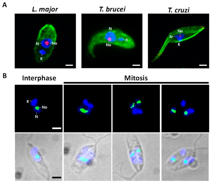
Interphase and mitotic nucleolus in trypanosomatids. (A) Fluorescence micrographs of L. major (procyclic promastigote stage), T. brucei (procyclic form), and T. cruzi (epimastigote stage). These three stages, which possess a single flagellum, grow and replicate in the corresponding insect host. They can be grown in large numbers in axenic culture media. These parasites have a single mitochondrion, which contains a network of thousands of catenated circular DNAs known as kinetoplast DNA. Parasites were fixed and treated with antibodies against nucleolar protein Nop56 from L. major (red) and α/β-tubulin (green) for visualization of the nucleolus and microtubules, respectively. Nuclear and kinetoplast DNA were counterstained with DAPI (blue). During interphase, the nucleolus is present as a single structure (red) located in a nucleoplasmic region weakly stained with DAPI. (B) Fluorescence images of L. major procyclic promastigotes during the cell cycle. Throughout the closed mitosis the nucleolus, represented here by Nop56, is conserved (green signal). During the course of the nuclear division, the round-shaped nucleolus is elongated and, eventually, split into two structures. Nuclear and kinetoplast DNA were counterstained with DAPI (blue). K, kinetoplast; N, nucleus; No, Nucleolus. Bar, 2 μm.

