Figure 4.
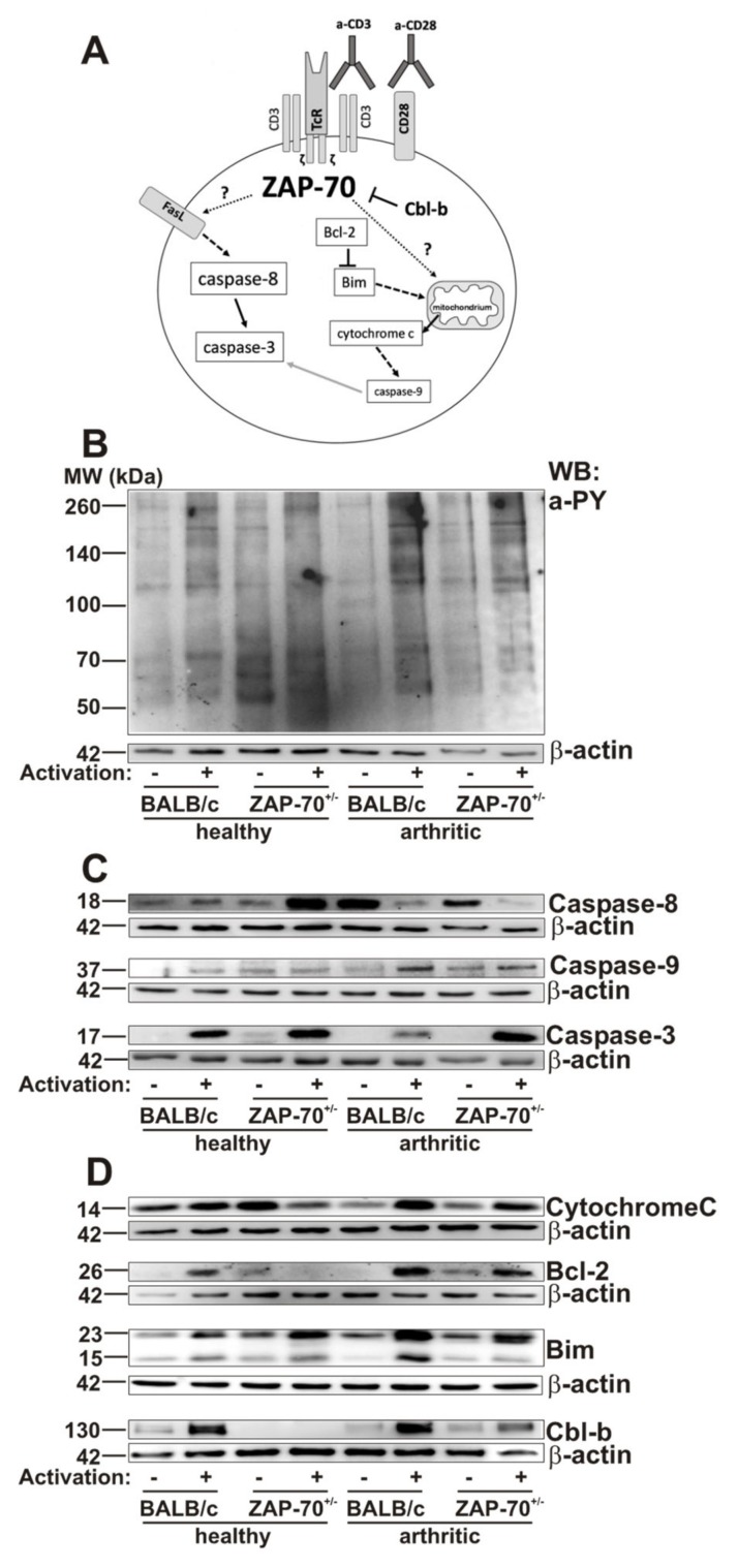
Phosphorylation patterns and expression of apoptotic proteins in healthy and arthritic mice were assessed with Western-blot. Schematic summary shows the studied activation and apoptosis pathways (A). T cells isolated from the spleens of healthy or arthritic BALB/c and ZAP-70+/− mice were lysed after 72 h of anti-CD3/anti-CD28 stimulation. Samples were separated using SDS-PAGE and detected by Western bloting using anti-phosphotyrosine (a-pY) (B), anti-Caspase-3, -8, and -9 (C), anti-Cytochrome C, anti-Bcl-2, anti-Bim or anti-Cbl-b (D) antibodies. Blots were reprobed with anti-β-actin–antibody to confirm equal sample loading. Figure shows representative blots from at least three independent experiments.

