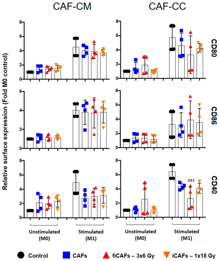
Figure 2.
Effects of CAFs on M1-macrophage cell surface markers. Monocyte-derived macrophages in non-stimulated or stimulated conditions were incubated with conditioned medium from irradiated (iCAF-CM) or non-irradiated CAFs (CAF-CM) or in (macrophage-CAF) co-cultures (CC). Resulting surface expression of M1-macrophage cell surface markers CD80, CD86 and CD40 were evaluated by flow cytometry. Filled black circles indicate surface levels in control macrophage cultures (M0 and M1). Results are expressed as fold M0-controls. Data represent mean (±SD) values from four-4 different CAF donors measured independently. Non-parametric Kruskal-Wallis test and p-values were determined between control and non-irradiated CAFs, control and the two iCAF-groups individually. iCAF (irradiated CAFs); fiCAFs: fractionated-irradiated CAFs.

