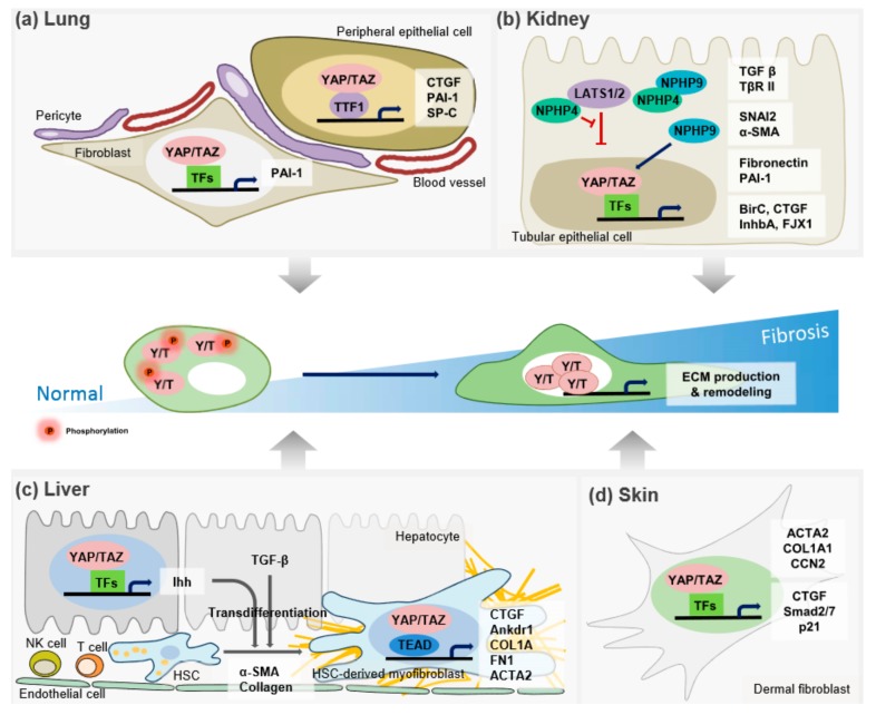
Figure 1.
The role of the Hippo pathway in the pathogenesis of tissue fibrosis. In normal cells, YAP/TAZ are phosphorylated and localized in the cytoplasm, leading to their inactivation. However, in many pathological situations, YAP/TAZ localize to the nucleus, and up-regulate their target genes to remodel the ECM. This action leads to tissue fibrosis. (a) In respiratory epithelial cells, binding YAP/TAZ with TTF-1 synergistically activates the expression of target genes, including SP-C, CTGF, and PAI-1. Also, active fibroblasts promote matrix synthesis via YAP/TAZ that are critical to the induction of profibrotic genes, such as CTGF and PAI-1, in the development of lung fibrosis. (b) NPHP4, by interacting with LATS1/2, promotes YAP/TAZ transcriptional activity. In contrast, NPHP9, another NPH proteins (NPHPs), directly interacts with YAP/TAZ and induces its nuclear localization to regulate a subset of target genes involved in renal fibrosis. (c) YAP/TAZ-mediated Indian hedgehog (Ihh) gene induction, which increases transcriptional reprogramming by modulating profibrotic gene expression in HSCs. In response to Ihh and TGF-β, HSCs transdifferentiate into the ECM-producing myofibroblasts. (d) YAP/TAZ are mainly localized with the nucleus, resulting in the profibrotic gene expression in dermal fibrolasts. See text for further details. The red spheres indicate the phosphorylation of YAP/TAZ by kinase.

