Figure 7.
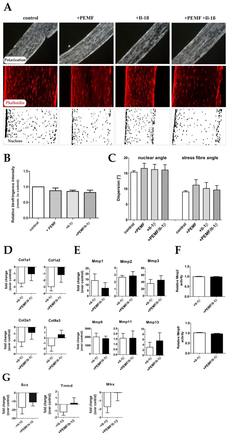
Extracellular matrix organization in tendon-like constructs before and after Il-1β and/or PEMF treatment. (A) Polarization microscopic images, phalloidin staining, and nuclear stain (DAPI) of 3D tendon constructs. (B) Relative birefringence intensities as surrogate for collagen fiber organization (n = 4). (C) Actin stress fiber angle and nuclear angle dispersion determined for the 4 treatment groups (n = 4). (D) The expression of extracellular matrix genes Col1a1, Col1a2, Col3a1 and Col9a3 is shows up-regulation PEMF treatment in pro-inflammatory stimulated (+PEMF +Il-1β) samples although not statistically significant (n = 9). (E) Under pro-inflammatory conditions the matrix metalloproteinases-1, -2, -3, -9, -11 and -13 show no significant changes in their expression after PEMF treatment (n = 7). (F) Relative MMP2 and MMP9 activity in Il-1β-stimulated constructs with and without PEMF exposure (n = 10). (G) Expression of tendon related marker genes Scleraxis (Scx), Tenomodulin (Tnmd), and Mohawk (Mkx) is restored after PEMF treatment of pro-inflammatory primed tendon constructs (n = 6).

