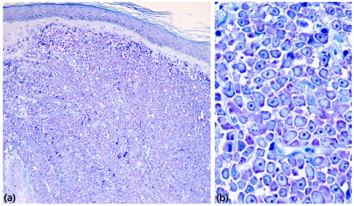
Figure 2.
Morphological findings in BPDCN. (a) Skin involvement, low-power Giemsa stain (original magnification 40×): a diffuse, monomorphous infiltrate massively involves the dermis, without epidermotropism. (b) The neoplastic cells are medium-sized blasts with fine chromatin and scanty cytoplasm, agranular on Giemsa staining (original magnification 600×).

