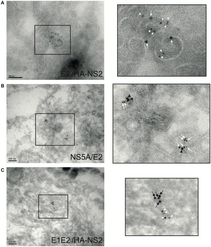
Figure 5.
Colocalization of HCV E2 glycoprotein, HCV E1E2 complex, NS2, and NS5A at the same ER membranes from the membranous web. A4HAHCVcc-infected cells were processed for electron cryomicroscopy double-labeling (A) E2 (AR3A + 10 nm gold-secondary Ab) and HA-NS2 (anti-HA + 6 nm gold-secondary Ab), (B) NS5A (9E10 + 10 nm gold-secondary Ab) and E2 (AR3A + 6 nm gold-secondary Ab), and (C) E1E2 complex (AR5A + 10 nm gold-secondary Ab) and HA-NS2 (anti-HA + 6 nm gold-secondary Ab). These representative images show the membranous web with E2 (white arrow), NS2 (white arrowhead), NS5A (black arrowhead), and E1E2 complex (black arrow) lying on the ER bilayer. The images in the column on the right are enlargements of the areas within the squares.

