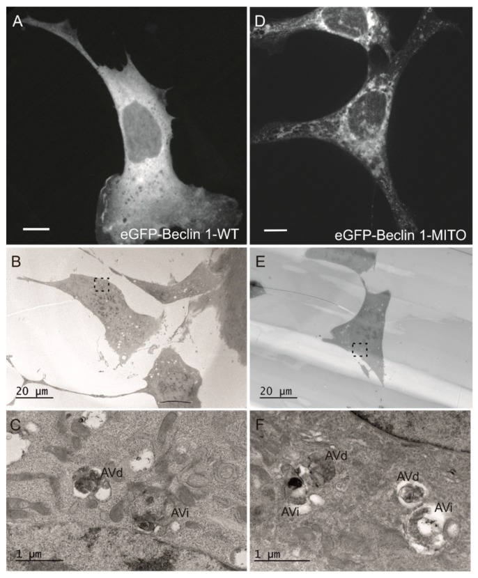
Figure 7.
Electron microscopy analysis of autophagosomes in MEF-ULK1/2-KO cells expressing eGFP-Beclin 1-WT or eGFP-Beclin 1-MITO. The cells were transfected with eGFP-Beclin 1 constructs and starved in amino-acid free medium for 1 h before fixation. (A–C) eGFP-Beclin 1-WT. (A) Confocal image of a cell expressing eGFP-Beclin 1-WT. Scale bar, 10 μm. (B) Electron micrograph of the same cell as in panel A. (C) Higher magnification of the boxed area in B, showing AVi (initial autophagic vacuoles/autophagosomes) and AVd (degradative autophagic vacuoles). (D–F) eGFP-Beclin 1-MITO. (D) Confocal image of a cell expressing eGFP-Beclin 1-MITO. Scale bar, 10 μm. (E) Electron micrograph of the same cell as in panel D. (F) Higher magnification of the boxed area in E, showing AVi and AVd.

