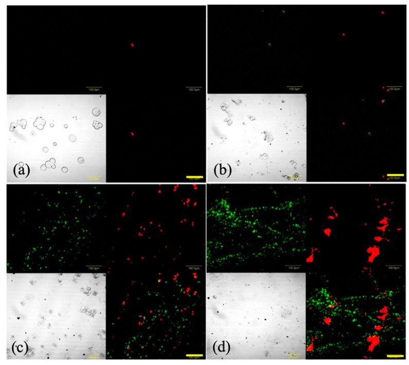
Figure 6.
Representative images of SNP cells stained with Annexin V-fluorescein isothiocyanate (green) and ethidium homodimer III (red) following photodynamic therapy (PDT). Cells were incubated with 8 μg/mL glucose-conjugated chlorin e6 (G-Ce6) for 24 h. Following washing with fresh medium, the cells were irradiated with 650-nm laser light (10 mW/cm2; 0, 1, 5, or 15 J/cm2). Following 4 h of PDT, the cells were stained using the Promokine Apoptotic/Necrotic Cells Detection kit. The images depict (a) 8 μg/mL G-Ce6 and 0 J/cm2 laser energy, (b) 8 μg/mL G-Ce6 and 1 J/cm2 laser energy, (c) 8 μg/mL G-Ce6 and 5 J/cm2 laser energy, and (d) 8 μg/mL G-Ce6 and 15 J/cm2 laser energy. Scale bar, 100 μm.

