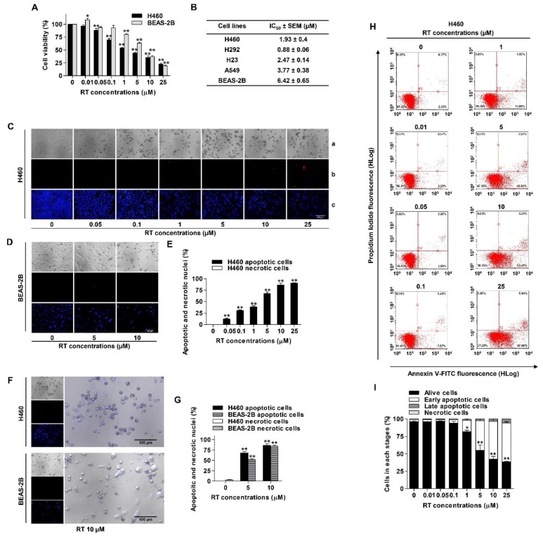
Figure 2.
Renieramycin T (RT) reduced cell viability and induced apoptosis in NSCLC and human normal lung epithelial (BEAS-2B) cell lines. (A) NSCLC and BEAS-2B cell lines were treated with various concentrations of RT (0–25 µM) for 24 h. Percentages of cell viability were determined using the MTT assay. (B) The half maximal inhibitory concentrations (IC50) in NSCLC and BEAS-2B cell lines were calculated by comparison with the untreated control. (C–G) H460 and BEAS-2B cell lines were treated with RT (0–25 µM) for 24 h. Hoechst 33342 and propidium iodide (PI) were added. Then, Images were detected by using an inverted fluorescence microscope (a–c). A condensed blue fluorescence of Hoechst 33342 reflected fragmented chromatin in apoptotic cells (c) while a red fluorescence of PI reflected late apoptotic or necrotic cells (b) comparing with no staining condition (a). Percentages of nuclear fragmented and PI positive cells were calculated. (H) H460 was treated with RT (0–25 µM) for 24 h. Apoptotic and necrotic cells were determined using annexin V-FITC/PI staining with flow cytometry. (I) Percentages of cells at each stage were calculated. Data represented the mean ± SEM (n = 3) (* 0.01 ≤ p < 0.05, ** p < 0.01, compared with the untreated control).

