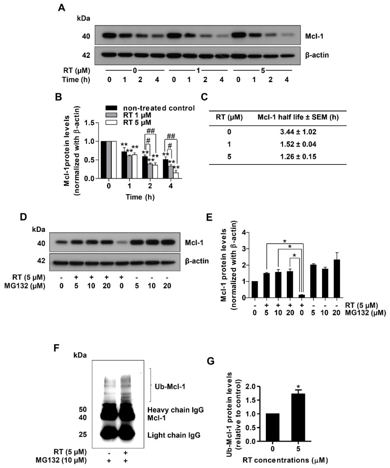
Figure 4.
Renieramycin T (RT) induced ubiquitin-mediated Mcl-1 proteasomal degradation. (A) Cycloheximide (CHX) chasing assay was performed to measure Mcl-1 half-lives. H460 cells were treated with RT (0–5 µM) with or without 50 µg/mL CHX as indicated by the time in h. Western blot analysis was performed for determined Mcl-1 levels. The blots were reprobed with β-actin to confirm equal loading of each of the protein samples. (B) The relative protein levels were calculated by densitometry (** p < 0.01, compared with the untreated control at 0 h, # 0.01 ≤ p < 0.05, ## p < 0.01, compared with the untreated control at the same time). (C) Mcl-1 half-lives were calculated. (D) H460 cell line was treated with RT (0–5 µM) with or without MG132 (0–20 µM) for 4 h. Mcl-1 expression levels were measured using Western blot analysis. The blots were reprobed with β-actin to confirm equal loading of each of the protein samples. (E) The reversal of RT-mediated down-regulation of Mcl-1 levels by MG132 was calculated by densitometry compared to the non-MG132 treated group (* 0.01 ≤ p < 0.05, compared with the non-MG132 treated group). (F) H460 was treated with RT (5 µM) and MG132 (10 µM) for 4 h. Then, protein lysates were collected subsequent to Mcl-1 immunoprecipitation, and the ubiquitinated protein levels were measured by Western blotting. (G) Ub-Mcl-1 levels were quantified using densitometry (* p < 0.01, compared with the untreated control) All data represented the mean ± SEM (n = 3).

