Figure 3.
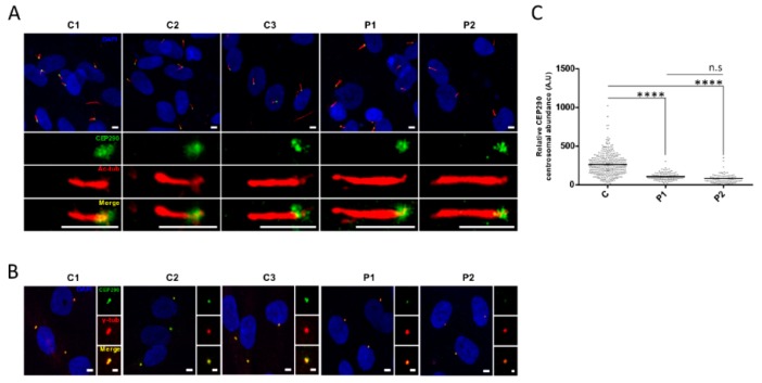
CEP290 expression assessment in quiescent cells. (A) Representative images of CEP290 (green) localization in quiescent control and mutant fibroblasts. Acetylated α-tubulin (Ac-tub; red) is used to stain the ciliary axoneme. As in control cell lines (C1–C3), CEP290 is correctly localized at the base of the cilia in patient (P1 and P2) fibroblasts. Scale bar, 5 µm. (B) Centrosomal localization of CEP290 (green) in control and mutant fibroblasts. The γ-tubulin (γ-tub; red) labeling is used as a centrosomal marker. Image scale bar, 5 µm and inset scale bar, 2 µm. (C) Quantification of the CEP290 immunofluorescence intensity at the basal body in each cell line (C represents the pooled values of C1–C3). Each dot depicts the labeling intensity of the protein in individual cells from six microscope fields (recorded automatically). The solid line indicates the mean. **** p ≤ 0.0001, n.s = not significant. A.U. = arbitrary unit.

