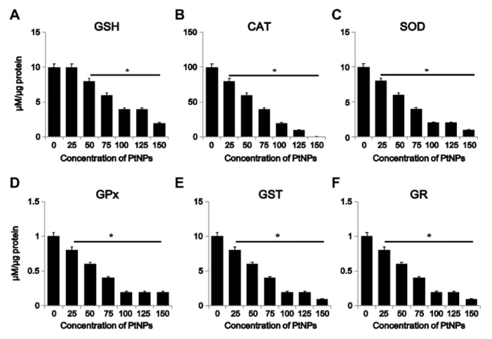
Figure 6.
Effect of PtNPs on anti-oxidant markers. THP-1 cells were treated with different concentrations of PtNPs (25–150 µg/mL) for 24 h. After incubation, the cells were harvested and washed twice with ice-cold phosphate-buffered saline solution. The cells were collected and disrupted by ultrasonication for 5 min on ice. (A) Glutathione (GSH) concentration was expressed as percentage of control. (B) Catalase (CAT) was expressed as percentage of control. (C) Superoxide dismutase (SOD) was expressed as percentage of control. (D) Glutathione peroxidase (GPx) concentration was expressed as percentage of control. (E) Glutathione S-transferase (GST) concentration was expressed as percentage of control. (F) Glutathione reductase (GR) concentration was expressed as percentage of control. Results are expressed as mean ± standard deviation of three independent experiments. There was a significant difference between treated and untreated cells per Student’s t-test (* p < 0.05).

