Figure 5.
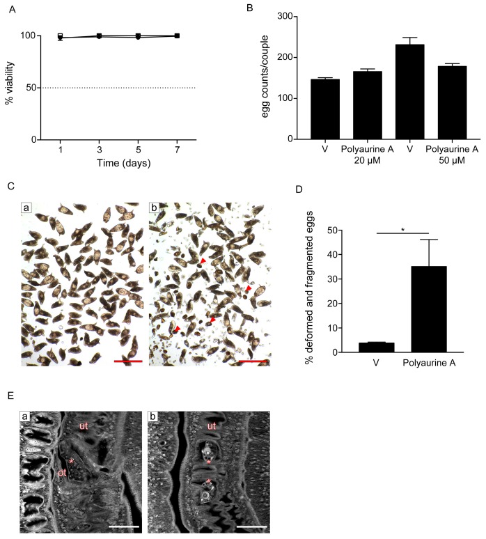
Effects of polyaurine A on S. mansoni adult parasites. (A) Adult pairs viability assays. Worm pairs were incubated with vehicle (DMSO) (circle), polyaurine A (1) 20 μM (square), or 50 μM (triangle), and viability is shown as the percentage of vehicle-treated samples indicated as 100%. The data shown represent the mean of three independent experiments ± SEM. (B) Worm pairs were incubated with vehicle (V) or polyaurine A (1) at the indicated concentrations, and eggs were counted at 72 h. Egg counts were normalized to the number of worm couples. The data shown represent the mean of three independent experiments ± SEM. (C) Representative microscopy images of S. mansoni eggs laid in vitro by worm pairs treated with vehicle (a) or 50 μM of polyaurine A (1) (b). Red arrows indicate the deformed or fragmented eggs (b). Scale bars = 200 μm. (D) Histograms represent the percentage of deformed and/or fragmented eggs counted at 72 h. Approximately 500 eggs were counted in 3 independent experiments (150–200 eggs/experiment). Asterisk in the figure indicates significant t-test p-value (* p < 0.05) relative to the comparison of 50 μM of polyaurine A-treated samples with the vehicle V-treated ones. (E) Representative confocal scanning laser images of adult S. mansoni pairs treated for 72 h with vehicle or 50 μM of polyaurine A. Eggs in the ootype (a, vehicle) or uterus (b, polyaurine A) are indicated by asterisks. Abbreviations: ot, ootype; ut, uterus. Scale-bars = 50 μm. In all experiments, vehicle-treated parasites or eggs received the same amount of DMSO (in volume) as the polyaurine A (1)-treated ones.

