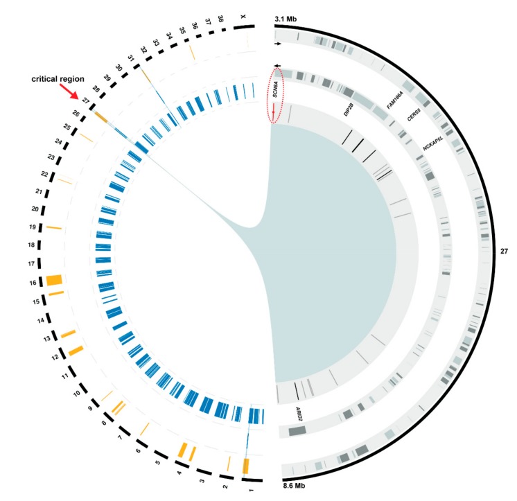
Figure 3.
Positional cloning of the spinocerebellar ataxia-associated variant. The canine chromosomes are depicted in the left half of the circle as black bars. Below, the 3 circular tracks from outside to inside indicate: (i) 23 genome segments from linkage analysis in orange, (ii) four homozygous blocks shared in affected dogs in blue, (iii) homozygous blocks found in unaffected dogs in blue. The initial four segments overlapping in cases are indicated by a blue background. Only a single interval on chromosome 27 showed both linkage and homozygosity in affected dogs and no homozygosity in unaffected dogs. Therefore, it was considered the critical region for spinocerebellar ataxia (red arrow). The right half of the circle displays a close-up view of this region (chr27:3,154,712-8,679,881) with 3 circular tracks below showing from outside to inside: (i, ii) gene content showing the 165 genes (grey boxes) annotated on both, the positive (outer circle) and negative (inner circle) strand, (iii) location of private intergenic (grey), intronic (black), and exonic (red) SNVs depicted as vertical lines. For clarity, only the names of six genes, in which an intronic or exonic SNV was found, are shown. Note the red ellipse highlighting the single protein-changing SNV in the SCN8A gene.

