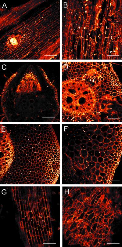
Figure 5.
Confocal laser scanning analysis of various organs in a 12-d-old double mutant. Longitudinal sections of a WT (A) and a double mutant (B) primary root. Cross sections of a WT (C) and a double mutant (D) primary root. Cross sections of a WT (E) and a double mutant (F) coleoptilar node. Longitudinal section of a young crown root from the coleoptilar node of a WT (G) and a double mutant (H). The positions of cross sections of the coleoptilar node were marked by arrows in Figure 4, C and F. Bars indicate 100 μm (C–F) or 200 μm (A, B, G, and H).

