Figure 5.
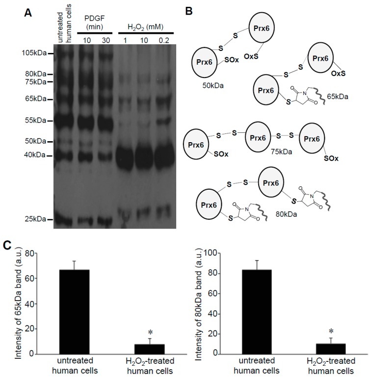
Identification of Prx6 multimers in human cells. (A) Human pulmonary artery smooth muscle cells were treated with or without PDGF (10 ng/mL) or H2O2 at indicated concentrations for 15 min. Cell lysates were prepared, incubated with Protein-SHifter Plus, subjected to SDS-PAGE without BME, and immunoblotted with the Prx6 antibody. (B) Possible structures of Prx6 that produce bands of various molecular weights. (C) Bar graphs represent the means ± SEM of the intensities of 65- and 80-kDa bands (n = 4). The symbol * represents the value significantly different from the untreated control value at p < 0.05.

