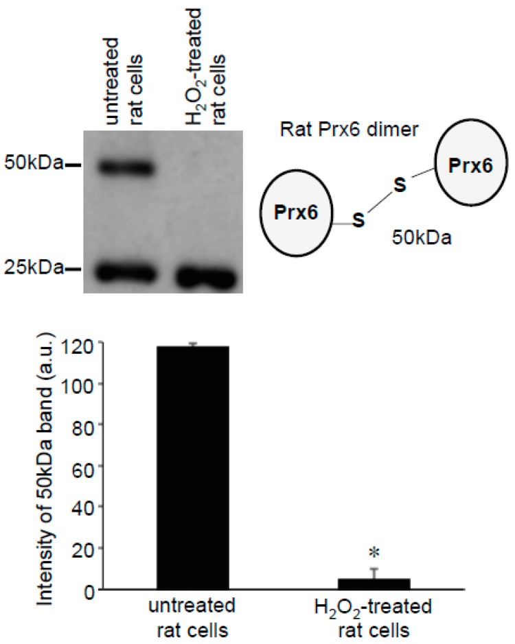
Figure 7.
Effects of H2O2 on Prx6 dimers in rat cells. Rat pulmonary artery smooth muscle cells were treated with H2O2 (200 μM) for 15 min. Cell lysates were prepared and subjected to SDS-PAGE and immunoblotting with the Prx6 antibody. No Protein-SHifters were added in these experiments. The bar graph represents the means ± SEM of the intensity of the 50-kDa band (n = 3). The symbol * represents the value significantly different from the untreated control value at p < 0.05.

