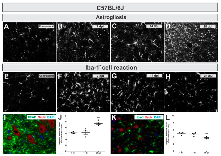
Figure 4.
Time-course of astrogliosis (A to D and I) and Iba-1+ cell reaction (E to H and L) in the lumbar spinal cord contralateral side and 7, 14 and 28 dpi post ventral root injury in WT mice. Note that astrogliosis increases over time. Contrarily, the Iba-1+ cell reaction is more intense in the acute phase after injury. I and K illustrate the glial reaction in the ipsilateral side nearby axotomized motoneuron-like cells, positive to NeuN (in red). (J and L) show the comparative quantification among time points post injury (ipsi/contralateral ratio); p < 0.001 (***). Scale bar = 50 µm.

