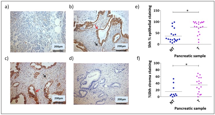
Figure A1.
Shh expression in normal and tumour pancreatic tissues. 19 paired normal and tumour primary pancreatic tissues and 1 unpaired tumour primary pancreatic tissue (39 total samples) were investigated for expression of the Hh pathway ligand, Shh by IHC analysis. Based on histophatological analysis the tumour tissues were divided into moderate (MDT) and poorly differentiated tumour (PDT). Representative images of Shh staining are shown: (a) normal pancreatic tissue; (b) moderately differentiated pancreatic tumour; (c) poorly differentiated pancreatic tumour; (d) negative control demonstrates absence of staining either in MDT tissue when an isotype control antibody was substituted for the specific antibody to the target. Red arrows and black arrows indicate epithelial and stromal cancer cells expressing Shh, respectively. Semi-quantitative analysis of the percentage Shh staining in the epithelial (e) and stromal (f) compartments in all tumour samples (* p < 0.05 Kruskal-Wallis multiple comparison Test).

