
Figure 3.
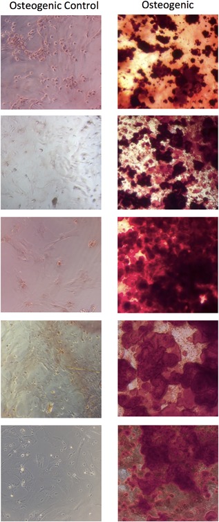
Light microscopy images (×10 magnification) of third passage cells from peripheral blood MSCs in rats treated with VEGF and AMD3100. The cells were cultured with osteogenic supplements for 21 days and stained with Alizarin red demonstrating mineral formation. Each row represents a culture from a different rat.