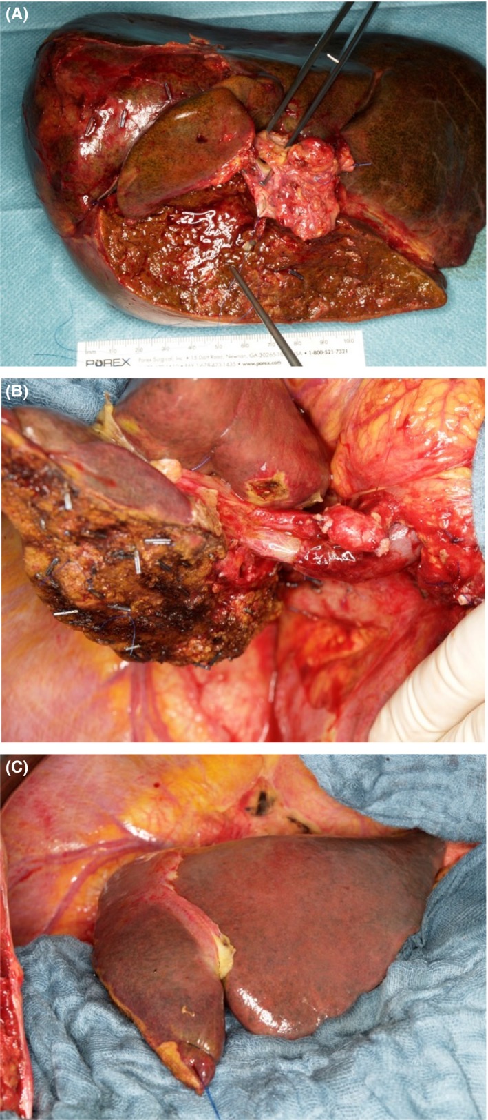
Figure 1.

(A) Resected specimen: extended right hemihepatectomy including segment I, extrahepatic bile duct, portal vein bifurcation and hilar tissue. Long suture at proximal cut end of left bile duct and forceps in resected portal vein bifurcation. (B) Lateral view of liver remnant (segments II, III and part of IV) after extended right hemihepatectomy with end‐to‐end anastomosis of the portal vein and transected left bile duct visible below left portal vein, prior to hepaticojejunostomy. (C) Anterior view of liver remnant (segments II, III and part of IV) after extended right hemihepatectomy
