
Figure 6.
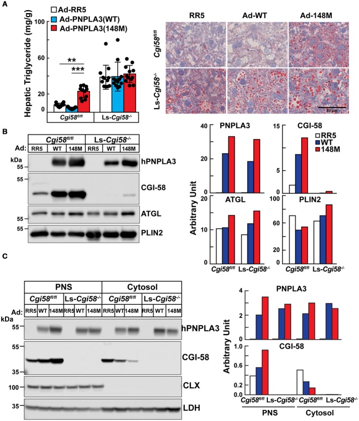
Hepatic lipid (A) and protein analyses (B,C) of Ls‐Cgi58‐/‐ mice infected with RR5, Ad‐PNPLA3(WT), or Ad‐PNPLA3(148M). (A) Chow‐fed Cgi58fl/fl and Ls‐Cgi58‐/‐ mice (n = 4/group, 8‐10 weeks old) were infected with recombinant Ad particles (1.25 × 10¹¹ genome copies) expressing no insert (Vector, RR5) or V5‐tagged versions of human PNPLA3(WT or 148M). Mice were killed 3 days later at the end of the last feeding cycle and hepatic TG levels were measured. Data were pooled from three independent experiments performed under similar conditions (A, left). Oil Red O staining was performed on liver sections at a magnification of ×20. Scale bar = 50 µm (A, right). (B) LDs were isolated from the mice described in (A), and the LD proteins were extracted and pooled. A total of 2 µg of LD protein was subjected to immunoblotting as described in the Materials and Methods. The signals were quantitated using LI‐COR and expressed in arbitrary units. (C) Proteins from the PNS and cytosol from the same mice were pooled and loaded onto the gel in equal proportions before immunoblotting, and the signals were quantitated using LI‐COR. The experiment was repeated twice and the results were similar. Abbreviations: CLX, calnexin; LDH, lactate dehydrogenase.