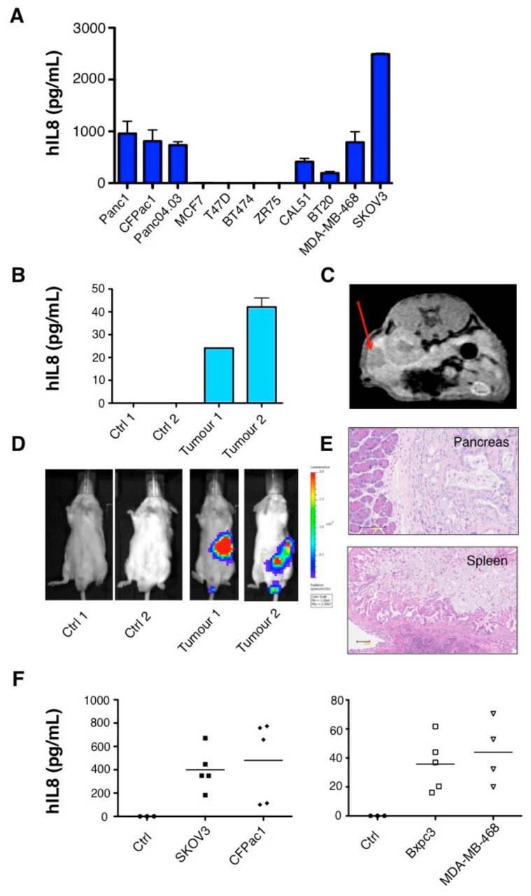
Figure 1.
Detection of Interleukin-8 (IL-8) in the supernatant of cancer cell lines and in the circulation of mice with solid tumor xenografts. (A) Supernatant was collected from pancreatic, luminal, and triple negative breast or ovarian cancer cell lines 48 h after seeding 5 × 10⁵ cells in a 6-well plate. Data show the mean ± standard error of the mean (SEM) of IL-8 measured using enzyme-linked immunosorbent assay (ELISA) in 3–4 independent experiments. (B) Plasma levels of human IL-8 in mice bearing orthotopic Panc04.03 pancreatic xenografts as detected by (C) magnetic resonance imaging (MRI) and (D) bioluminescence imaging (BLI). Red arrow denotes location of tumor in MRI scan in (C). Control (Ctrl) mice were tumor-free. (E) Orthotopic tumor infiltration of pancreas and spleen was confirmed histologically by H and E staining of tissue. Scale bar: 100 µM. (F) Circulating human IL-8 was detected in the plasma of mice bearing intraperitoneal tumor xenografts of ovarian, pancreatic, and breast origin. Data show the mean and individual data points for each mouse.

