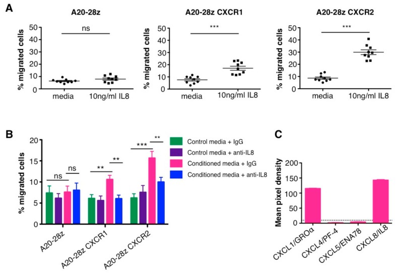
Figure 4.
In vitro migration of chemokine receptor CAR T-cells to IL-8. (A) Media or media containing 10 ng/mL recombinant human IL-8 was added to the lower well of a transwell migration plate. CAR T-cells were added to the upper chamber and the percentage of migrated T-cells in the lower well was quantified after 2 h. Data show the percentage of migrated cells in triplicate wells from 3 independent experiments (mean ± SEM); *** p < 0.0001, calculated using Student’s t-test. (B) Conditioned media from SKOV3 cells that contained either an IgG control or an anti-IL-8 blocking antibody was added to the lower well of a transwell migration plate. CAR T-cells were added to the upper chamber and the percentage of migrated T-cells in the lower well was quantified after 2 h. Data show the percentage of migrated cells within triplicate wells from 3 independent experiments (mean ± SEM); ns: not significant; ** p < 0.01; *** p = 0.0001 calculated using an unpaired, two-tailed Student’s t-test. (C) Secretion of CXCL1 and IL-8 (CXCL8) by SKOV3 cells. Supernatant from SKOV3 was applied to nitrocellulose membranes spotted with capture antibodies and then detected using biotinylated antibodies followed by chemiluminescent detection reagents. Mean pixel density from duplicate wells was calculated using Image J software.

