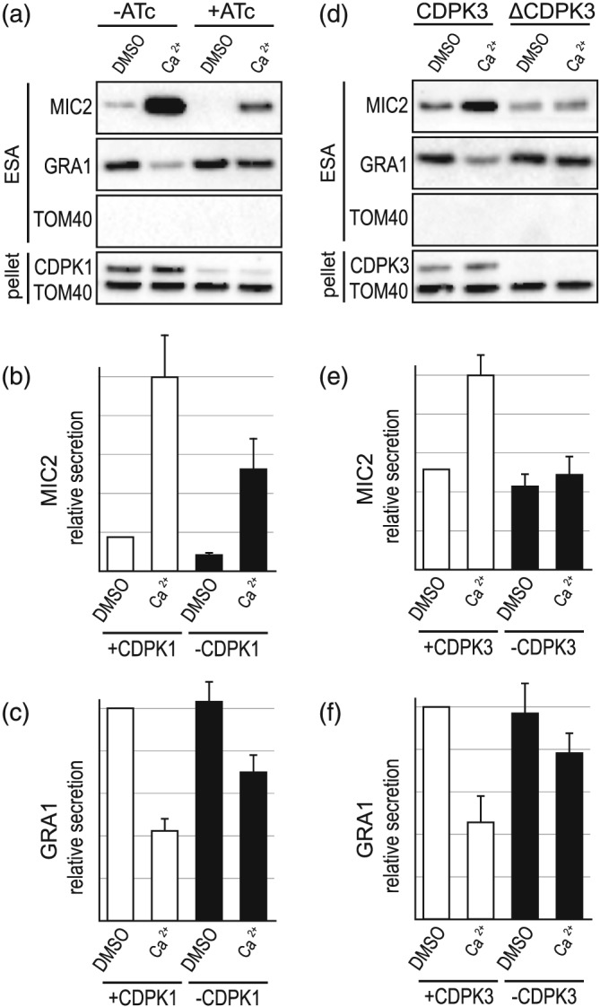
Figure 3.

Effect of loss of CDPK1 or CDPK3 on Ca2+‐induced microneme protein (MIC) and granule protein (GRA) secretion. (a–c) Extracellular secretion assays (ESA) with and without ATc‐induced CDPK1 depletion in iΔHA‐CDPK1 cells. Depletion of HACDPK1 is seen by immuno‐detection of HA in the cell pellet. A23187 (5μM) is used for Ca2+ stimulation. Relative secretion of MIC2 (b) and GRA1 (c) is shown (n = 8). (d–f) ESA and relative secretion (n = 7) measurements for wildtype (+CDPK3) versus CDPK3 knockout (−CDPK3) cells. Absence of CDPK3 is seen by CDPK3 immuno‐detection in the cell pellet (d). TOM40 in ESA serves a control for cell lysis and controls for cell equivalent loading in the pellet. Error bars = SEM
