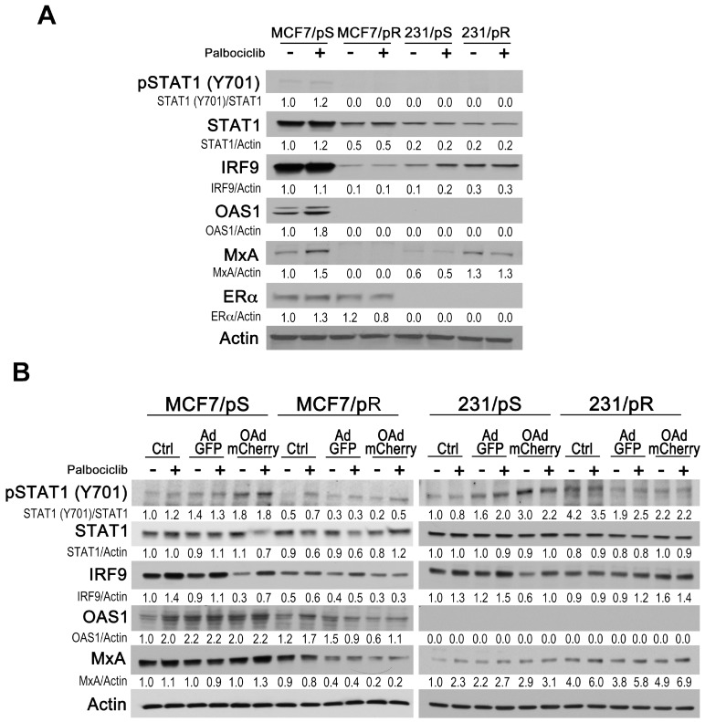
Figure 5.
Type I IFN response in ER+ and ER− breast cancer cells. (A) Cells were treated with either the vehicle (H2O) or palbociclib (500 nM), harvested after 24 h, and immunoblotted with the indicated antibodies. (B) Cells were either left uninfected (Ctrl) or infected with AdGFP or OAdmCherry at a multiplicity of infection (MOI) concentration of five alone or in combination with 500 nM palbociclib for 24 h. Type I IFN signaling was assessed by immunoblotting with the indicated antibodies. Quantitative densitometry analysis is shown relative to loading control normalized to palbociclib-sensitive control-treated cells.

