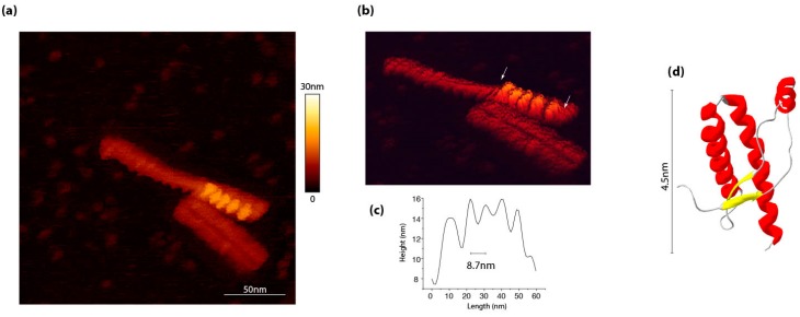
Figure 2.
Motif repetition along PrPSc assemblies, as identified by atomic force microscopy. (a) 263K PrPSc assemblies purified according to a protocol by Wenborn et al. [52], as observed by atomic force microscopy in a liquid environment in sodium acetate buffer pH 5.0. (b) The scanning performed by using an Olympus AC406 nm cantilever in a QI mod revealed the existence of periodic element indicated between arrows in panel. (c) The axial distance between each periodical element is around 8.7 nm. (d) For comparison, the globular domain of ovine recombinant PrP (PDB:1TPX) has approximatively a diameter of 4.5 nm.

