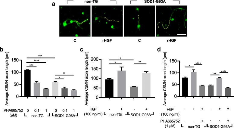
Fig. 5.
Treatment with rHGF enhanced the axonal outgrowth of CSMNs in motor cortical cultures. a-d SOD1-G93A TG mice were sacrificed at P3. After dissociating motor cortices, 20,000 cells were seeded on 24-well plates. Three days later, cells were fixed and subjected to immunocytochemistry (ICC) assay. a Representative image of CSMNs. Antibodies specific to UCHL1 (green) and Ctip2 (red, not shown) were used to label CSMNs. Serum-free media was used as a control medium (C). Cells were visualized with confocal laser scanning microscopy. The axon length of CSMNs was measured using Fiji software (yellow line). The axon length of CSMNs was measured after treatment with PHA665752, an inhibitor for Met (b) or rHGF (c) or rHGF plus PHA665752 (d). For bar graphs, values are represented as mean ± SEM. In Fig. 5b-d, one-way ANOVA was performed, followed by Tukey’s post-hoc test. *p < 0.05, **p < 0.01, ***p < 0.001, ****p < 0.0001. Scale bar: a = 50 μm

