
Figure 2.
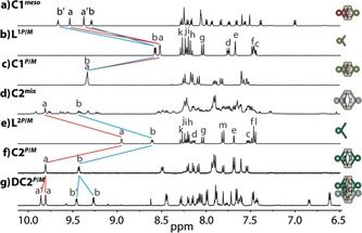
1H NMR spectra (400 MHz, [D6]DMSO, 293 K) of: a) C1 meso, b) L1P/M, c) homochiral C1 P/M, d) a statistical mixture of C2 stereoisomers, e) L2P/M, f) homochiral C2 P/M, and g) the homochiral interpenetrated cage structure DC2 P/M (here: 600 MHz, [D3]acetonitrile, 293 K).