Figure 2.

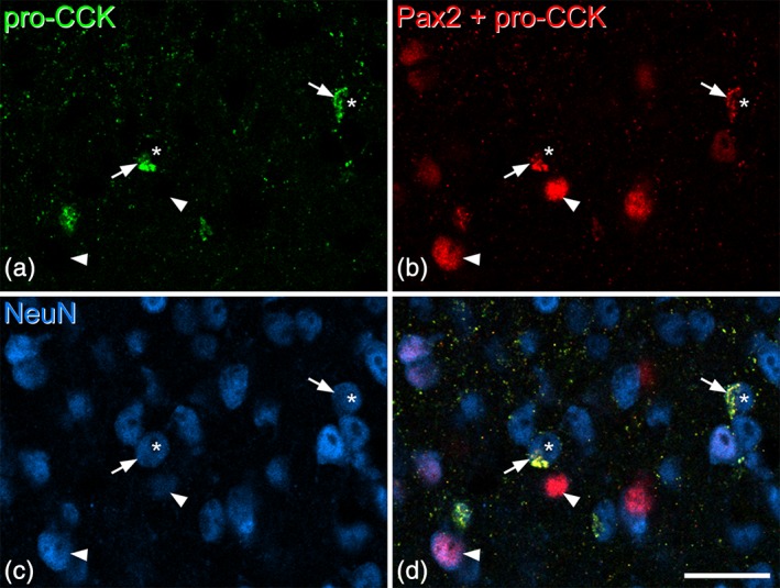
Lack of Pax2 immunoreactivity in the nuclei of pro‐cholecystokinin (CCK)‐positive cells. These images are from a section that had been reacted with rabbit pro‐CCK antibody (revealed with Alexa 488) and mouse NeuN antibody (revealed with Alex 647). The section was subsequently reacted with Pax2 antibody (also raised in rabbit), which was revealed with Rhodamine Red. (a) Staining in the Alexa 488 channel is restricted to pro‐CCK‐positive structures, which represent perikaryal cytoplasm of CCK‐expressing neurons. (b) Staining in the Rhodamine channel detects both pro‐CCK and Pax2, because the Rhodamine‐conjugated secondary anti‐rabbit antibody was applied after both rabbit primary antibodies (pro‐CCK and Pax2). However, these can readily be distinguished because the Pax2 profiles are not labeled with Alexa 488 and because Pax2 staining is nuclear, whereas pro‐CCK is cytoplasmic. (c) The same field scanned to reveal NeuN. (d) The merged image shows pro‐CCK‐immunoreactivity in the perikaryal cytoplasm of cells (two marked with arrows) that have nuclei that are negative for Pax2 (asterisks). Two Pax2‐positive cells, which lack pro‐CCK, are indicated with arrowheads. All images are from a single optical section. Scale bar = 20 μm [Color figure can be viewed at wileyonlinelibrary.com]
