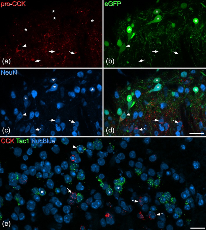
Figure 6.

Limited coexpression of cholecystokinin (CCK) and substance P in the dorsal horn. (a–d) Part of the superficial dorsal horn from a Tac1Cre mouse that had been injected with AAV.flex.eGFP 8 days prior to perfusion fixation. (a–c) Show immunostaining for pro‐CCK, enhanced green fluorescent protein (eGFP) fluorescence, and NeuN immunoreactivity, while (d) shows a merged image. Numerous eGFP‐positive (substance P‐expressing) cells can be seen in the upper part of the field (which corresponds to laminae I‐II), and most of these lack pro‐CCK (some are indicated with asterisks). There are several pro‐CCK‐positive (eGFP‐negative) cells in the lower part of the field, some marked with arrows. A double‐labeled cell is marked with an arrowhead. Images are from four optical sections at 1 μm z‐spacing. (e) A single confocal optical plane from a section that had been reacted with probes against CCK and Tac1 mRNAs, and counterstained with NucBlue. The field of view contains numerous Tac1 mRNA‐positive cells (three marked with asterisks), which form a dense band across the middle of lamina II. There are also several CCK mRNA‐positive cells (three shown with arrows), and most of these are located more ventrally. A single cell with both Tac1 and CCK mRNAs is indicated with an arrowhead. Scale bars for (a–d) and for (e) = 20 μm [Color figure can be viewed at wileyonlinelibrary.com]
