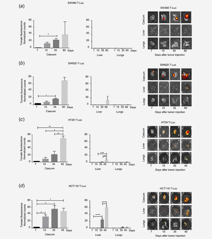
Figure 3.

Different metastatic potential of the four human colon carcinoma cell lines implanted in SCID beige mice. Primary tumors (caecum) and organs with metastases (liver, lungs) were harvested from SW480 T‐Luc (a), SW620 T‐Luc (b), HT29 T‐Luc (c) and HCT116 T‐Luc (d) tumor‐bearing mice at various cancer development time‐points. Counts of TdTomato fluorescence were normalized on in vitro quantification for each cell line (n = 3–4 mice, average ± S.E.M.). Representative fluorescence imaging of one mouse per group is shown. ****p < 0.0001, ***p < 0.0005, **p < 0.005, *p < 0.05.
