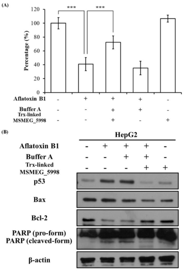
Figure 4.
The effect of AFB1 and Trx-linked MSMEG_5998 on the viability and apoptosis of HepG2 cells. (A) Cell viability was determined by using MTT assay. Before the assay, 1 × 10⁴ cells/well in 96-well plates were treated, or not, with AFB1, buffer A, and Trx-linked MSMEG_5998 for 48 h. ***, p < 0.001. Each experiment was replicated three times. (B) The levels of p53 and apoptosis-related proteins were detected by western blotting in HepG2 cells after the cells had been treated as described in (A). β-actin was used as an internal control. Because of the similar molecular weights of the analyzed proteins, the proteins were analyzed on separate SDS-PAGE gels.

