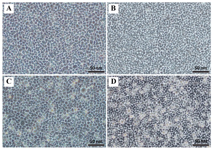
Figure 1.
Microscopic examination of epithelioma papulosum cyprini cells infected with European chub iridovirus. (A) Control flask at 48 h post-inoculation (hpi); (B) control flask 96 hpi; (C) infected flask showing enlarged and refractile cells at 48 hpi; (D) infected flask showing enlarged and refractile cells at 96 hpi. Scale bars are 50 µm.

