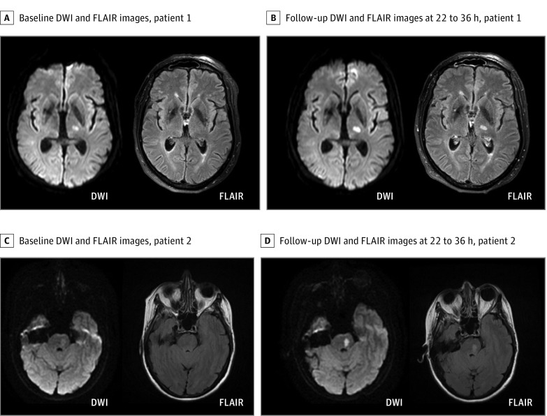
Figure 1. Magnetic Resonance Imaging in Patients With Lacunar Infarcts.
Example magnetic resonance images of 2 randomized patients with a lacunar infarct in the left internal capsule (A) and brain stem (C). Before randomization for treatment (A and C), both infarcts presented with a hyperintense signal on diffusion-weighted imaging (DWI) without signal alterations in the corresponding area on fluid-attenuated inversion recovery (FLAIR). According to the mandatory DWI-FLAIR mismatch, patients were randomized and treated either with alteplase or placebo. Follow-up magnetic resonance images 22 to 36 hours after treatment (B and D) also show a hyperintense signal on FLAIR corresponding to the lesion area detected on DWI.

