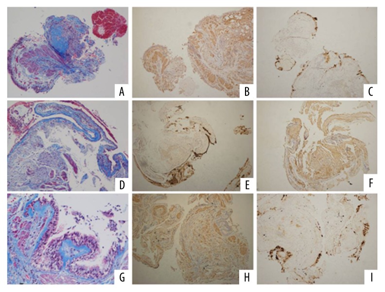
Figure 2.
Pathology features in the patients with IPF, the lung tissue was obtained through transbronchial lung biopsy. [(A) Masson staining (collagen fiber) showed disorder alveolar structure and interstitial fibrosis in IPF patients (×100/HP); (B) α-SAM (fibrinogen specific antibody) staining (×100/HP); (C) The characteristic pathological changes of terminal alveolar bronchopathy with submucosal gland structure in terminal alveolar structure. MUC5B stain was positive by immunohistochemistry assay (×100/HP); (D) Masson staining (collagen fiber) showed that the alveolar structure was disorder and interstitial fibrosis was seen in IPF patients; (E) α-SAM (fibrinogen specific antibody) staining (×100/HP); (F) Terminal alveolar bronchopathy with submucosal gland structure with MUC5B stain positive (×100/HP); (G) Masson staining (×200/HP); (H) α-SAM (fibrinogen specific antibody) staining (×100/HP); (I) MUC5B positive expression (×100/HP)].

