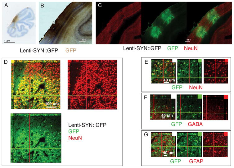
Figure 4. Lentivirus synapsin promoter expression in visual cortex.
A–C. Coronal sections of V1 injected with Lenti-SYN::GFP. A,B. GFP visualized with HRP-DAB staining. C. Tiled confocal scan of section stained for GFP and NeuN. Same section in all images with GFP visualized in green (DyLight 488) and NeuN in red (Alexa 568). D–E. Confocal slice to show dense expression of GFP in NeuN positive neurons. Note the sharp transition border of viral GFP expression. F. GFP does co-localized with GABA positive interneurons (red) G. GFP does not co-localized with GFAP positive glia (red).

