
Figure 3: CD8+ T-cells are required for anti-tumor immunity induced by CHK1i with or without anti-PD-L1 blockade.
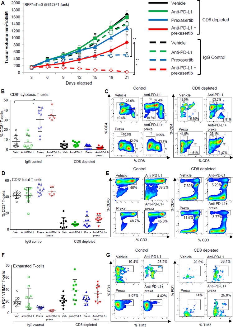
(A) Tumor growth curves +/−SEM from vehicle, prexasertib alone (10mg/kg, 2 out of 7 days, BID), anti-PD-L1 alone (300μg, 1 out of 7 days) and prexasertib+anti-PD-L1 treatment groups in RPP B6 mice in IgG control and CD8-depleted (anti-CD8, 200μg, 2 out of 7 days) groups.
(B-C) CD8+ T cells measured by flow cytometric analysis in single-cell suspensions prepared from tumors (n=10) in CD8-depleted groups as compared to IgG control groups. The analysis was independently repeated at least three times. t-test, p<0.0001.
(D-H) SCLC tumors in Fig 3A were harvested at Day 21 and the immune profiling was analyzed by FACS at the endpoint, the representative plots and cumulative data for all the tumors is shown. FACS analysis of CD3+CD45+ Total T-cells (D-E), exhausted CD8 T cells: CD45+CD3+CD8+PD-1+TIM3+ (F-G) from the endpoint primary tumors. The statistical summary is shown with ANOVA test. ns, no significance; *, p < 0.05; **, p < 0.001; ***, p < 0.0001.