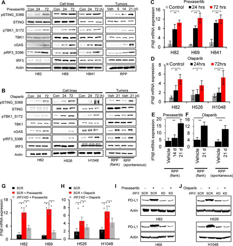
Figure 5: Anti-tumor immune response post-DDR targeting is mediated via STING-TBK1-IRF3 pathway in SCLC.

(A-B) Immunoblots of markers in the STING pathway including total and phospho STING (S366), total and phospho TBK1 (S172), cGAS, total and phospho IRF3 (S396) in lysates collected from SCLC cell lines and tumors treated with prexasertib (A) or olaparib (B). Actin served as a loading control.
(C-D) Quantitative PCR (qPCR) measurement of IFNβ mRNA expression in SCLC cell lines 24 and 72 hours after prexasertib (C) and olaparib (D) treatment.
(E-F) Quantitative PCR (qPCR) measurement of IFNβ mRNA expression in SCLC tumor models after treatment with prexasertib in RPP flank model (E) and olaparib in RPP flank and spontaneous (F) SCLC in vivo models.
(G-H) qPCR measurement of IFNβ mRNA expression 72 hours after treatment with prexasertib (G) and olaparib (H). IRF3 expression was knocked down 24 hours prior to drug treatment using siRNA targeting IRF3. A scrambled siRNA control (SCR) is included. All data representative of mean ± SD and p values represented as * p<0.05, **p<0.001, ***p<0.0001.
(I-J) Immunoblot of PD-L1 in SCLC cells treated with prexasertib (I) and olaparib (J) (72 hours). IRF3 expression was knocked down in all cells 24 hours prior to drug treatment using siRNA targeting IRF3. A scrambled siRNA control (SCR) is included.
