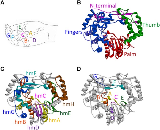
FIGURE 3.

Domains, motifs, and homomorphs of a typical calicivirus RdRp. (A) Representation of a slightly cupped right hand resembling an RdRp with the position of motifs A to G on fingers, palm, and thumb. (B–D) Ribbon diagrams of the RHDV RdRp (PDB ID: 1KHW); (B) fingers, palm, and thumb domains colored blue, red, and green, respectively, and the N-terminal domain colored magenta; (C) structurally conserved homomorphs (hmA to hmH); and (D) functional motifs A to G (the positions of homomorphs and corresponding motifs are indicated by the same color). Ribbon diagrams were generated using Discovery Studio (Dassault Systèmes BIOVIA, Discovery Studio Visualizer v17.2.0, San Diego: Dassault Systèmes, 2016).
