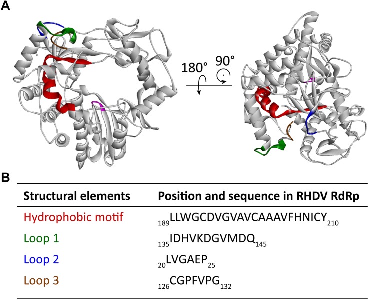
FIGURE 5.
Localization of a partially buried hydrophobic membrane interaction motif in the RHDV RdRp. (A) Ribbon diagrams of the RHDV RdRp (PDB ID: 1KHW). The hydrophobic motif is colored red, loop 1 green, hydrophobic loop 2 blue, and hydrophobic loop 3 brown. The active site (motif C) is highlighted magenta to provide a reference point for the position of the hydrophobic motif in the RdRp. (B) Amino acid positions and sequences of the structural elements highlighted in the diagrams above. Ribbon diagrams were generated using Discovery Studio (Dassault Systèmes BIOVIA, Discovery Studio Visualizer v17.2.0).

