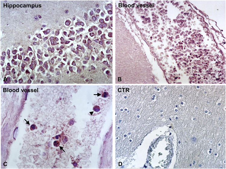
Figure 4. Immunohistochemical localization of TIMP1 in DKA brains.
TIMP1 was localized by indirect immunoperoxidase in DKA brains as described in Materials and Methods section. Hippocampal neurons exhibit low TIMP1 staining (A). In addition, reduced staining of TIMP1 was found in cells present in the blood vessels (B, C). Some intravascular TIMP1+ cells had morphology typical for neutrophils (C, arrows), while others for lymphocytes (C, arrowhead). Controls of Immunoperoxidase were negative (D). Magnification: x400 (in A, C and D) and x200 (in B).

