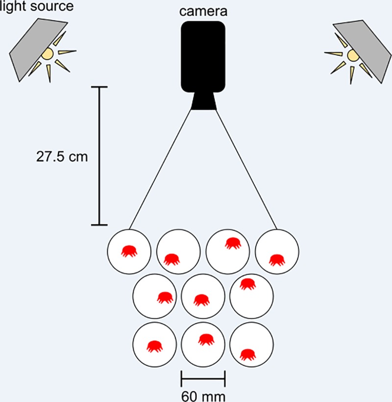
Fig 1. Experimental setup.

A camera was mounted 27 cm directly above a collection of 10 petri dishes, with one varroa mite per dish. Two utility lamps were mounted in a square surrounding the recording area, angled to minimize glare off the dish and maximize illumination of the recording area (figure not to scale). Once the footage was recorded, mites were individually transferred to micropipette tubes and placed in a -80°C freezer for subsequent analysis of viral loads.
