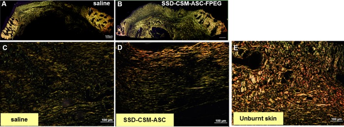
Fig 9. Maturation of collagen.
Picrosirius staining on day 28 infected burn wounds treated with (A) saline or (B) SSD-CSM-ASC-FPEG. Zoomed images from A shows (C) pre-dominantly thinner (green) regrowing fibers in infected saline control samples, (D) more number of thicker, arranged mature collagen fibers (red) in wounds treated with SSD-CSM-ASC-FPEG, and a basket like mature collagen in the (E) unburnt skin.

